

Your images, your formats
- DICOM, PNG, JPEG and TIFF accepted directly
- Multi-slice DICOM studies, greyscale or RGB, with slice navigation
- Format, integrity and OCT-modality validated on upload
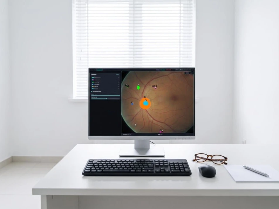
Automated analysis of OCT and fundus images that returns segmented biomarkers, a classified diagnosis and a ready structured report — in under a minute, in your PACS or in our cloud.

Every study in one list: type, clinician, status, whether biomarkers were found. A new one is added by dropping a file — DICOM or an image: .dcm, .png, .jpg, .jpeg, .bmp, .tiff.
Open the platform
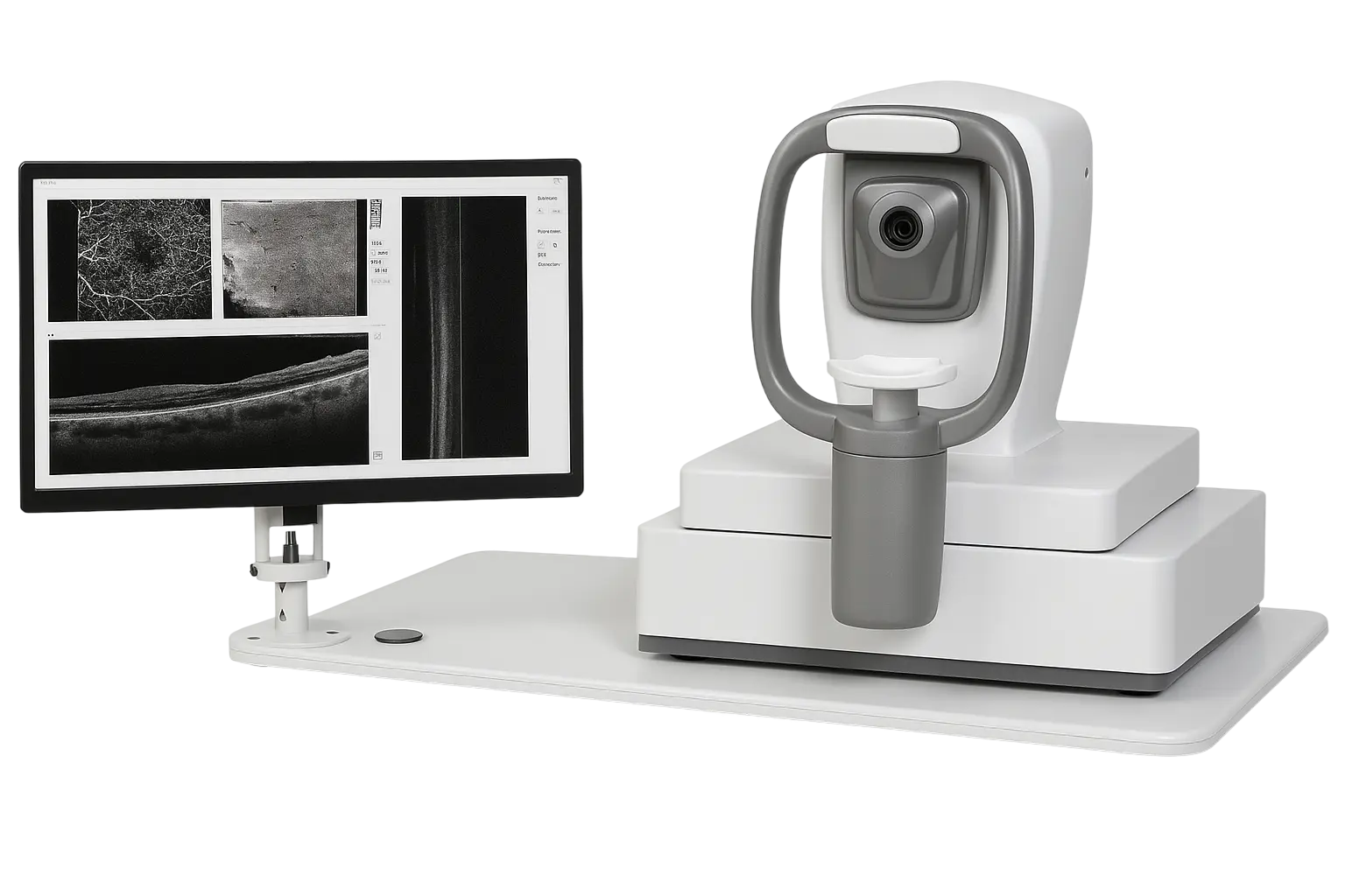

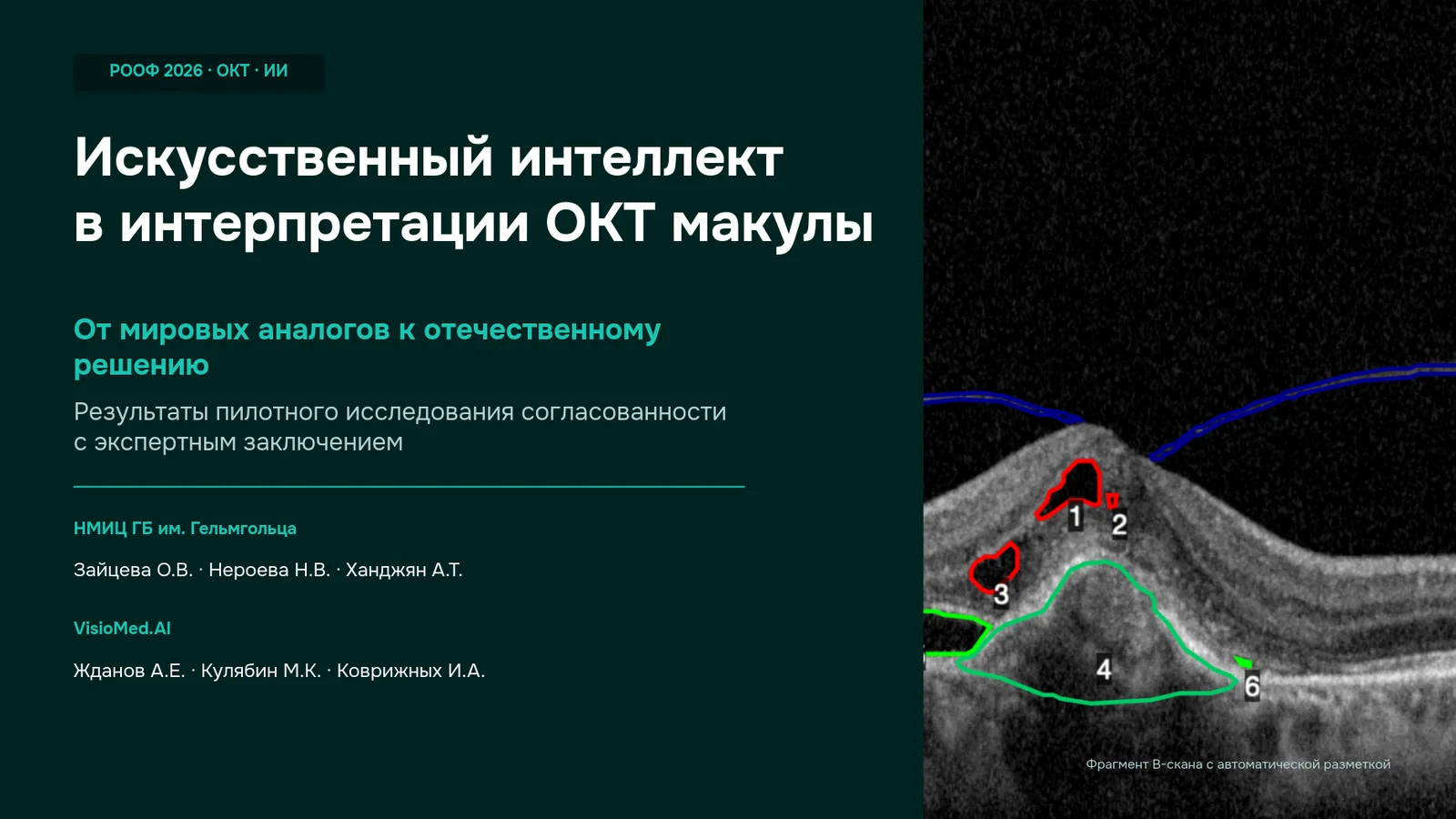
Biomarker segmentation, classification and a finished report for an OCT or fundus image. The clinician gets layers and prose that only need checking and signing.
Predicting outcome from a series of scans: response to anti-VEGF therapy and the choice between injections and surgery. The value is in the trajectory, not in one image.
A second opinion and an assistant: it searches a patient's studies, cross-references the findings and drafts the conclusion.
The VisioMed.AI solution is not only a different approach to diagnosing eye diseases but also a significant reduction in the doctor's routine duties, which together allows us to focus on what's most important – our patients and their health.

A trained neural network can be a good assistant for quick diagnosis. But most importantly, it is useful in the face of an impending staffing shortage. Let me explain: tomographs are available in both private and public clinics. But only a small number of ophthalmologists specializing in retinal diseases can work with and understand the images.

As part of the international research group 'Retinalbiomarkers', we see enormous potential for AI in ophthalmology. The use of AI in OCT analysis not only improves diagnosis but also automates the detection of pathologies, increases the accuracy and speed of image interpretation, leading to more effective patient treatment.
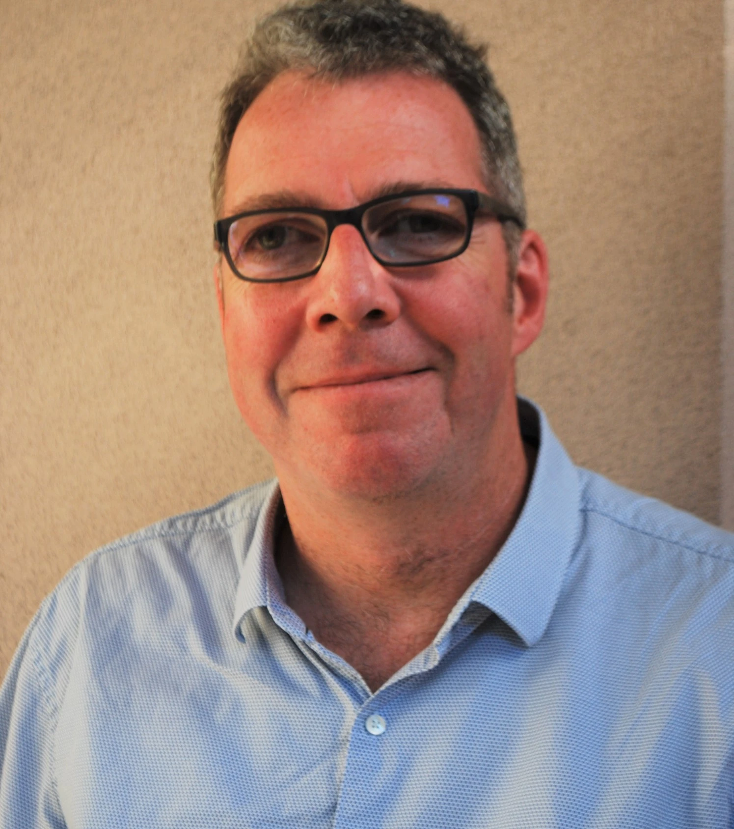
Together with VisioMed.AI, we are developing artificial intelligence for ophthalmosurgeons to improve diagnostic accuracy and better treatment strategy selection for macular holes and other retinal diseases. This solution minimizes subjective errors, automates image analysis, and accelerates clinical decision-making.
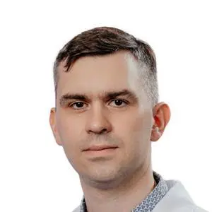
During the development of VisioMed.AI solutions, doctors manually train artificial intelligence algorithms, which allows AI to adopt expert knowledge and adapt to various clinical scenarios. This approach provides an opportunity to get a second opinion on the presence of a disease, reduces the likelihood of diagnostic errors, and increases the objectivity of OCT image analysis.
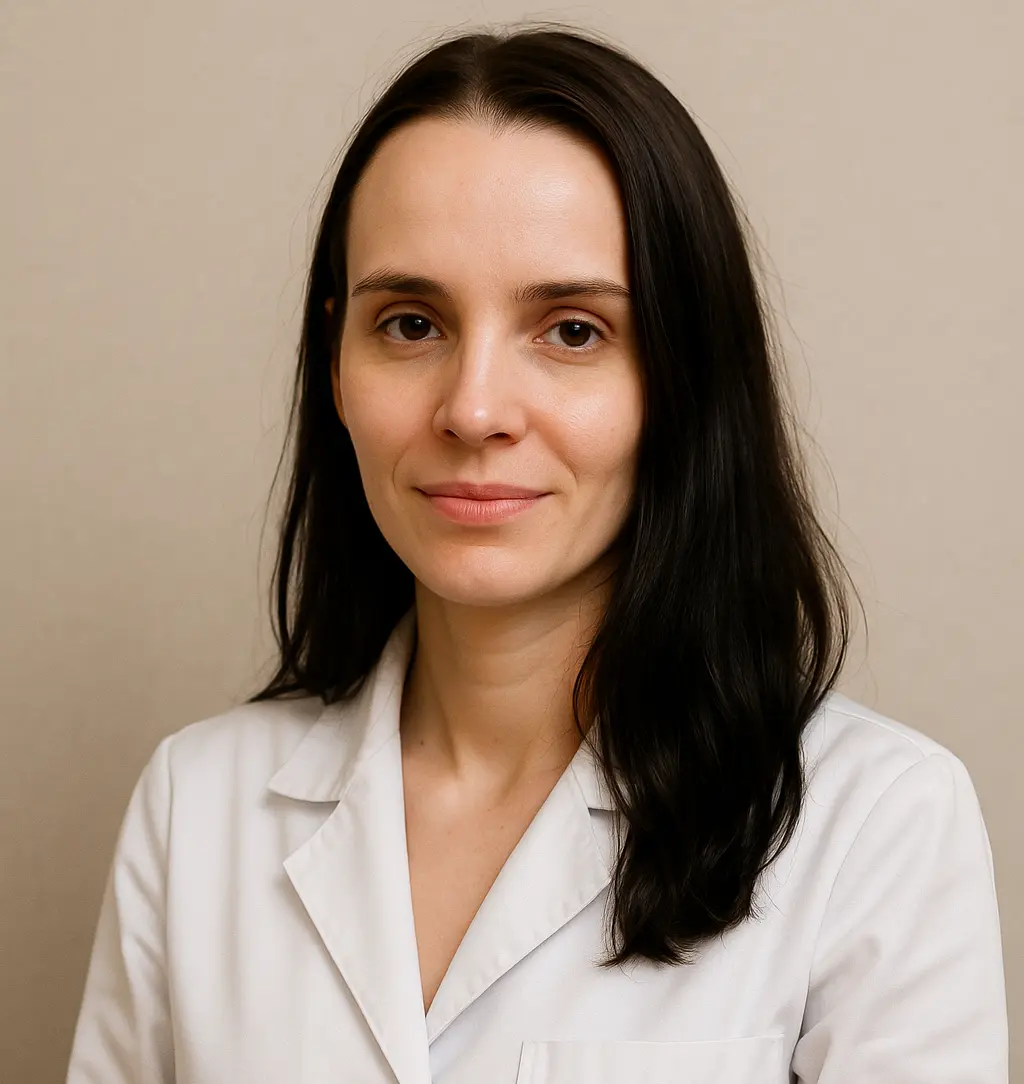
The VisioMed.AI solution will become an indispensable tool for novice ophthalmologists, helping them to better understand the structure and pathologies of OCT images. It is not just a diagnostic tool but also a powerful educational platform that contributes to the development of young specialists' professional skills.

Tell us what you image and we will set up a pilot on your data — in our cloud, or entirely inside your network.